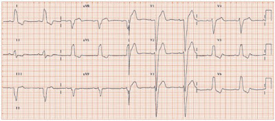
Left Bundle Branch Block (LBBB).
In left bundle branch block there is a delay or blockage of conduction in the main left bundle branch. This affects the normal depolarisation in the ventricles.
In left bundle branch block, the impulse begins in the SA node and depolarises the atria, then travels through the AV node to the left and right bundle branches. On finding the left bundle branch blocked, the impulse travels down the right bundle branch.
The septum is unable to depolarise from left to right as normal and instead depolarises from right to left. If V1 is our electrode, the impulse will initially be travelling away from it, as the septum depolarises. This causes a negative deflection (Q wave) to be inscribed. However, this is often not visible. Right ventricular depolarisation follows. If VI is our electrode, the impulse would now travel towards the electrode so a small positive deflection (R wave) would be inscribed. The left ventricle is then depolarised. The impulse is now travelling away from V1 and so a negative deflection (S wave) would be inscribed. Thus, the pattern of left bundle branch block in V1 is negative.
As V6 sits opposite V1, it will be affected in the opposite way. Therefore the small Q waves that are usually seen in the lateral leads will not be evident. The entire QRS complex in left bundle branch block will be widened (greater than 0.12 seconds) owing to the conduction delay.
The most common causes of LBBB:
- Coronary artery disease,
- Hypertension,
- Cardiomyopathy.
- Lev disease (sclerosis of the cardiac skeleton),
- Lenegere disease (primary degenerative disease of the conduction system),
- Advanced rheumatic heart disease, calcific aortic stenosis.
- An iatrogenic cause of LBBB is produced during cardiac pacing, because the pacing wire usually abuts the right ventricle and induces an LBBB-like morphology on the ECG.
ECG criteria for LBBB:

- QRS duration > 0.12 seconds,
- a broad monomorphic R wave in leads I, V5, and V6,
- a wide S wave following an initial small (or absent) R wave in the right precordial leads,
- absence of septal Q waves in leads I, V5, and V6.
The term incomplete LBBB is used to describe these findings in patients who have a QRS complex
duration < 0.12 seconds
Read Also: Right Bundle Branch Block


