Posts

Showing posts with the label Acute Pulmonary Embolism

Mechanical Complications of Venous Catheterization and their Countermeasures
Image
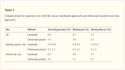
  Arterial puncture and hematoma Central venous catheterization rarely accompanies the formation of hematoma. However, with the jugular vein, and particularly when the carotid artery is mistakenly punctured, the formation of a hematoma may block the upper airway. If an artery is mistakenly punctured during SV puncture, external compression to stop the bleeding can be difficult to apply. If a catheter of 7 Fr (equivalent to a diameter of 2.3 mm) or less is inserted into an area, where compression is possible and the catheter can be withdrawn and external compression applied for 10 min, then it can be withdrawn with no problem. In other words, if an artery is mistakenly punctured by a needle thinner than 14 G (equivalent to a diameter of 2.1 mm), hemostasis may be possible via compression. However, if a catheter or dilator larger than 7 Fr is inserted into an artery or a vessel for which compression is not possible, a cardiovascular surgeon should be brought ...

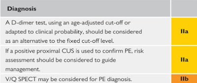
D-dimer Test. Elevated D-dimer level. D-dimer Range in Pragnancy

Image
D-dimer is a breakdown product of fibrin protein, which is present in the blood after the destruction of a blood clot. The products are formed during the lysis of a blood clot under the influence of plasmin and some fibrinolytics. In a number of pathological conditions characterized by the activation of blood coagulation, a constant process of formation of a fibrin clot occurs in the body, and then its splitting. 

Clinical presentation of Acute Pulmonary Embolism

Image
The clinical signs and symptoms of acute PE are non-specific. In most cases, Pulmonary Embolism  is suspected in a patient with dyspnoea, chest pain, pre-syncope or syncope, or haemoptysis. Haemodynamic instability is a rare but important form of clinical presentation, as it indicates central or extensive PE with severely reduced haemodynamic reserve. Syncope may occur, and is associated with a higher prevalence of haemodynamic instability and RV dysfunction.  Conversely, and according to the results of a recent study, acute PE may be a frequent finding in patients presenting with syncope (17%), even in the presence of an alternative explanation.