Posts

Showing posts with the label respiratory

Extubation Strategies after Cardiac Surgery

Image
Extubation Strategies after Cardiac Surgery  

Assessing Patient Readiness for Extubation

Image
  Assessing Patient Readiness for Extubation

Spontaneous Breathing Trial

 Chris Nickson Spontaneous breathing trials (SBT) are used to identify patients who are likely to fail liberation from mechanical ventilation SBT is “the defacto litmus test for determining readiness to breathe without a ventilator” Ideally, during an SBT we want to observe the patient under  conditions of respiratory load that would simulate those following extubation

Cuff Leak Test

Image
Cuff Leak Test    Chris Nickson OVERVIEW The cuff leak test is used to predict risk of post-extubation stridor in intubated patients Use and interpretation of the test needs to take into account the overall context of the patient’s condition and the management implications does not exclude obstruction due to supraglottic structures being splinted apart while the ETT is in situ PROCEDURE Suction endotracheal and oral secretions and set the ventilator in the assist control mode with the patient receiving volume-cycled ventilation With the cuff inflated, record displayed inspiratory and expiratory tidal volumes to see whether these are similar Deflate the cuff Directly record the expiratory tidal volume over the next six breathing cycles as the expiratory tidal volume will reach a plateau value after a few cycles Average the three lowest values The difference between the inspiratory tidal volume (measured before the cuff was deflated) and the averaged expiratory tidal volume (...

Cardiogenic Pulmonary Edema

Image
Cardiogenic Pulmonary Edema keywords: cardiogenic pulmonary edema, heart failure, pulmonary congestion, acute dyspnea, left ventricular dysfunction, fluid overload, cardiac edema      Increased hydrostatic pressure in the pulmonary capillaries results in increased transvascular fluid filtration and is most often caused by volume overload or impaired left ventricular function resulting in elevated pulmonary vascular pressures.

Indications for Intubation and Mechanical Ventilation.

Cardiorespiratory arrest or impending arrest.

Airway Device Selection.

Image
Face Mask LMA ETT Procedure duration Short Short–Medium Any Protection against aspiration No No Yes Peak inspiratory pressure* < 20 cmH2O < 20 cmH2O Any Stimulation by device Low Medium High LMA Laryngeal Mask Airway ETT Endotracheal Tube * Pressures above 20 cmH2O may distend the stomach

Pulmonary Function and Perioperative Mortality/Morbidity.

Image
    Increased morbidity results from hypoxemia, hypoventilation resulting in acute hypercarbia, pulmonary infection, prolonged intubation, and mechanical ventilation. Patients presenting for lobectomy or pneumonectomy as well as those with pulmonary comorbidity must have pulmonary function and arterial blood gas values. A forced expiratory volume in 1 second (FEV1) less than 800 ml in a 70-kg person is probably incompatible with life and is an absolute contraindication to lung resection because of the high incidence for extended mechanical ventilation.

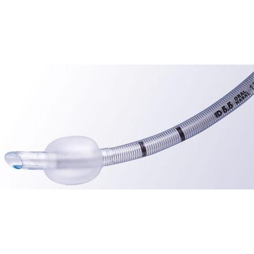
Appropriate endotracheal tube sizes and approximate depths.

Image
         Endotracheal tube sizing:

Pathological Breathing Patterns.

Image

Steps for successful insertion of a laryngeal mask.

Image
The laryngeal mask airway (LMA) is an acceptable alternative to mask anesthesia in the operating room. It is often used for short procedures when endotracheal intubation is not necessary.   Here some important steps for successful insertion of LMA   (click to enlarge) : Relative contraindications for the LMA: patients with pharyngeal pathology (eg, abscess) pharyngeal obstruction full stomachs (eg, pregnancy,hiatal hernia) low pulmonary compliance (eg, restrictive airways disease) requiring peak inspiratory pressures greater than 30 cm H2O.