Posts

Showing posts with the label Guidelines

ICU Management Post-Cardiac Surgery 2022

Image
  Webinars 2022-Management of post-operative cardiac surgery patients in ICU

GUIDELINES FOR INSERTION OF IMPLANTABLE CARDIOVERTER-DEFIBRILLATORS

Image
AMERICAN COLLEGE OF CARDIOLOGY, AMERICAN HEART ASSOCIATION, AND HEART RHYTHM SOCIETY GUIDELINES FOR INSERTION OF IMPLANTABLE CARDIOVERTER-DEFIBRILLATORS Class I Heart Failure:

Difficult Airway Decision Pathways

Image
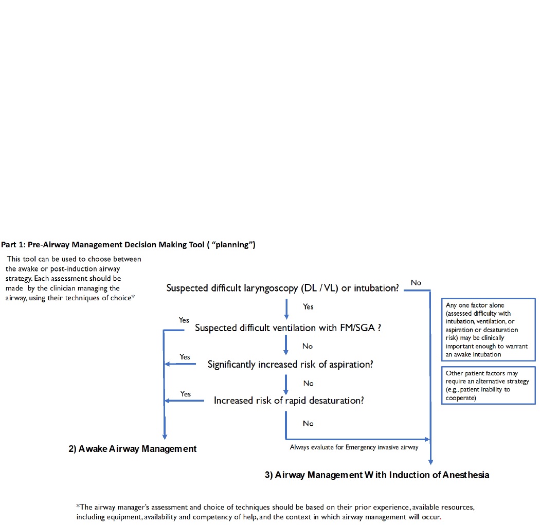
Difficult Airway Decision Pathways   Part 1 is a decision tool that incorporates relevant elements of evaluation and is intended to assist in the decision to enter the Awake Airway Management or Airway Management with the Induction of Anesthesia arms of the ASA difficult airway algorithm.

Anaesthesia and caring for patients during the COVID-19 outbreak. Airway Management Guidelines.

Image
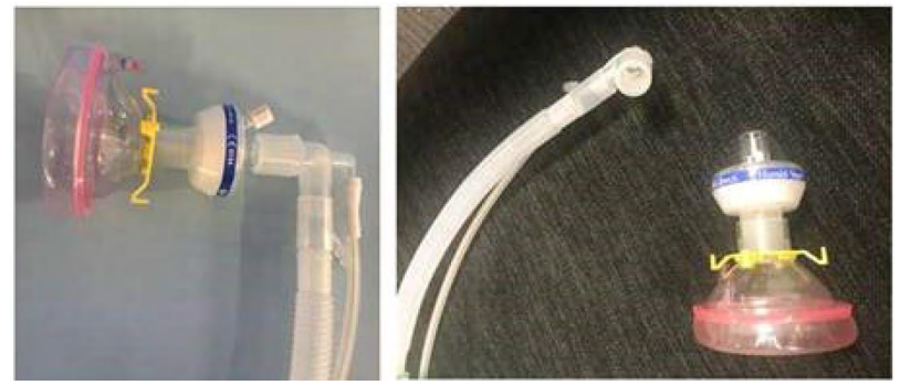
  AIM  To advise on processes and techniques required to protect staff and prevent transmission of infection during airway procedures for patients with known or suspected COVID-19 infection. To help anaesthesia staff and anaesthesia departments in clinical decision making and in the production of local processes and protocols. This advice is not intended to replace officially produced local advice (where available) from hospital departments, health services or state or federal health bodies.

Cautionary notes specific to pediatric central venous catheterization

Image
Caution during puncture Ultrasound-guided techniques are recommended for children with small veins that are difficult to puncture. Select a thinner, lighter probe than for adults. A linear probe or hockey-stick probe can be used. Recommended catheters

Mechanical Complications of Venous Catheterization and their Countermeasures

Image
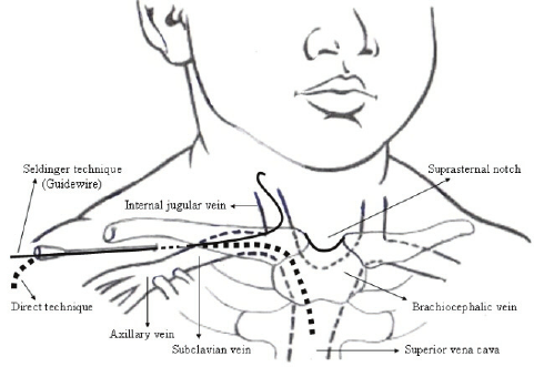
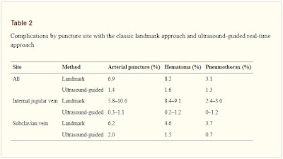
  Arterial puncture and hematoma Central venous catheterization rarely accompanies the formation of hematoma. However, with the jugular vein, and particularly when the carotid artery is mistakenly punctured, the formation of a hematoma may block the upper airway. If an artery is mistakenly punctured during SV puncture, external compression to stop the bleeding can be difficult to apply. If a catheter of 7 Fr (equivalent to a diameter of 2.3 mm) or less is inserted into an area, where compression is possible and the catheter can be withdrawn and external compression applied for 10 min, then it can be withdrawn with no problem. In other words, if an artery is mistakenly punctured by a needle thinner than 14 G (equivalent to a diameter of 2.1 mm), hemostasis may be possible via compression. However, if a catheter or dilator larger than 7 Fr is inserted into an artery or a vessel for which compression is not possible, a cardiovascular surgeon should be brought ...

Confirmation Methods for Central Venous Catheterization

Image
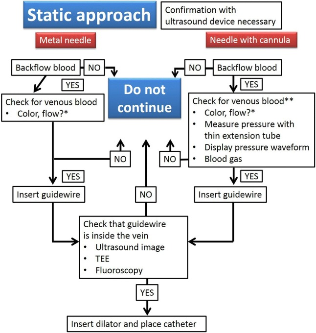
  The spread of ultrasound devices has reduced the frequency of mechanical complications. However, critical situations can arise when mechanical complications occur. In a 2004 report, the central venous catheter-related mechanical complications with the highest mortality rates were pulmonary artery damage, hemothorax, cardiac tamponade , and air embolism, in that order .

Selection of Intravenous Catheters and Sites

Image
  Peripheral and Midline Catheter Recommendations 1. In adults, use an upper-extremity site for catheter insertion. Replace a catheter inserted in a lower extremity site to an upper extremity site as soon as possible. Category II  2. In pediatric patients, the upper or lower extremities or the scalp (in neonates or young infants) can be used as the catheter insertion site. Category II 

Optimal Central Venous Catheter Placement

Image
  Confirm that the catheter is placed correctly in the vein and that the tip is in the desired position using a chest radiograph. Ideally, the tip of the catheter should be roughly parallel with the wall of the superior vena cava, caudal to the inferior margin of the clavicle, between the third rib and the fourth/fifth thoracic vertebra, and cranial to the bifurcation of the trachea or right primary bronchus (see Fig  ).

Chemical Thromboprophylaxis after Cardiac Surgery

Image

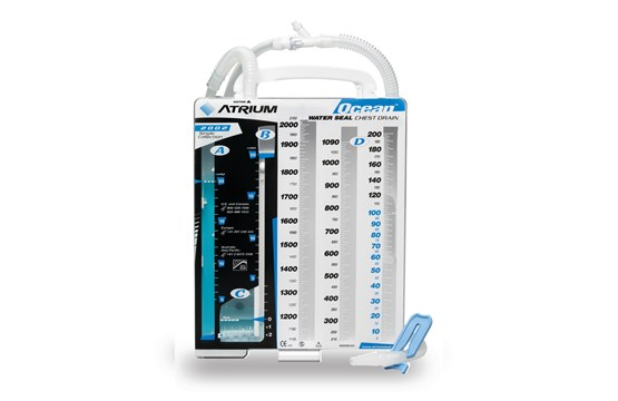
Chest Tube Patency in Cardiac Surgery

Image
  Chest Tube Patency keywords: chest tube patency, cardiac surgery, retained blood syndrome, postoperative complications, atrial fibrillation, chest tube clogging, active tube clearance, pericardial drainage, mediastinal blood, tamponade prevention, heart surgery recovery, chest tube management, anticoagulation therapy, pleural effusion, surgical drainage

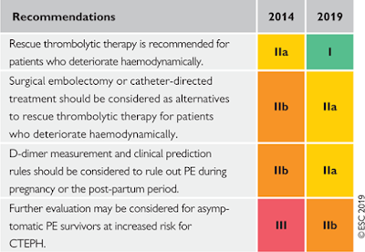
Acute Pulmonary Embolism. Changes in recommendations 2014–19

Image
What is new in the 2019 Guidelines?

Practice Guidelines for Central Venous Access: A Report by the American Society of Anesthesiologists Task Force on Central Venous Access

Image
What other guideline statements are available on this topic? Several major organizations have produced practice guidelines on central venous access  128   –   132   Why was this Guideline developed? The ASA has created this new Practice Guideline to provide updated recommendations on some issues

Practice Advisory for Intraoperative Awareness and Brain Function Monitoring: A Report by the American Society of Anesthesiologists Task Force on Intraoperative Awareness

Image
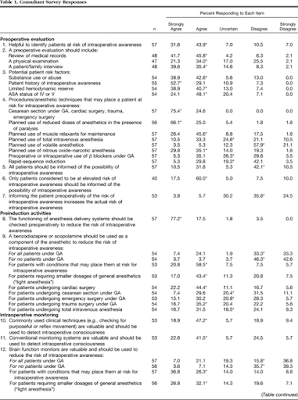
PRACTICE advisories   are systematically developed reports that are intended to assist decision making in areas of patient care. Advisories provide a synthesis and analysis of expert opinion, clinical feasibility data, open forum commentary, and consensus surveys. Advisories are not intended as standards, guidelines, or absolute requirements. They may be adopted, modified, or rejected according to clinical needs and constraints.

Practice Guidelines for Acute Pain Management in the Perioperative Setting: An Updated Report by the American Society of Anesthesiologists Task Force on Acute Pain Management

Image
What other guideline statements are available on this topic? These Practice Guidelines update the “Practice Guidelines for Acute Pain Management in the Perioperative Setting,” adopted by the ASA in 2003 and published in 2004.* Why was this guideline developed? In October 2010, the Committee on Standards and Practice Parameters elected to collect new evidence to determine whether recommendations in the existing Practice Guideline were supported by current evidence. How does this statement differ from existing guidelines? New evidence presented includes an updated evaluation of scientific literature and findings from surveys of experts and randomly selected ASA members. The new findings did not necessitate a change in recommendations. Why does this statement differ from existing guidelines? The ASA guidelines differ from the existing guidelines because they provide new evidence obtained from recent scientific literature as well as findings from new surveys...

Management of Dyslipidemias 2019, Lipid Guidelines

Image
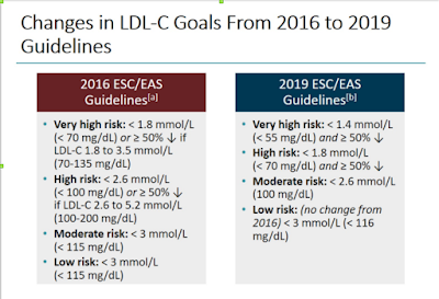
The following are key points to remember from the 2019 European Society of Cardiology (ESC)/European Atherosclerosis Society (EAS) guidelines for the management of dyslipidemias: lipid modification to reduce cardiovascular (CV) risk:

The Glasgow coma scale (GCS) in practice.

Image
         The Glasgow coma scale (GCS) gives a reliable, objective way of recording the conscious state of a person. It has value in predicting ultimate outcome. 3 types of response are assessed, note in each  case the best response (or best of any limb) which should be recorded.

Intravenous fluid therapy. Routine maintenance. NICE guideline.

Image
Routine maintenance 1.4.1 If patients need IV fluids for routine maintenance alone, restrict the initial prescription to: 25–30 ml/kg/day of water  and

American Society of Anesthesiologists Recommendations for Preanesthesia Checkout Procedures

Image
To Be Completed Daily. Item 1: Verify that auxiliary oxygen cylinder and self-inflating manual ventilation device are available and functioning. Item 2: Verify that patient suction is adequate to clear the airway.

Stable Angina or Silent Ischaemia: Indications for Revascularisation 2019

Image
Stable Angina also known as 'effort angina', this refers to the classic type of angina related to myocardial ischemia.