Posts

Showing posts with the label catheterization

Cautionary notes specific to pediatric central venous catheterization

Image
Caution during puncture Ultrasound-guided techniques are recommended for children with small veins that are difficult to puncture. Select a thinner, lighter probe than for adults. A linear probe or hockey-stick probe can be used. Recommended catheters

Mechanical Complications of Venous Catheterization and their Countermeasures

Image
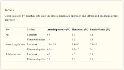
  Arterial puncture and hematoma Central venous catheterization rarely accompanies the formation of hematoma. However, with the jugular vein, and particularly when the carotid artery is mistakenly punctured, the formation of a hematoma may block the upper airway. If an artery is mistakenly punctured during SV puncture, external compression to stop the bleeding can be difficult to apply. If a catheter of 7 Fr (equivalent to a diameter of 2.3 mm) or less is inserted into an area, where compression is possible and the catheter can be withdrawn and external compression applied for 10 min, then it can be withdrawn with no problem. In other words, if an artery is mistakenly punctured by a needle thinner than 14 G (equivalent to a diameter of 2.1 mm), hemostasis may be possible via compression. However, if a catheter or dilator larger than 7 Fr is inserted into an artery or a vessel for which compression is not possible, a cardiovascular surgeon should be brought ...
Selection of Intravenous Catheters and Sites

Image
  Peripheral and Midline Catheter Recommendations 1. In adults, use an upper-extremity site for catheter insertion. Replace a catheter inserted in a lower extremity site to an upper extremity site as soon as possible. Category II  2. In pediatric patients, the upper or lower extremities or the scalp (in neonates or young infants) can be used as the catheter insertion site. Category II 

Optimal Central Venous Catheter Placement

Image
  Confirm that the catheter is placed correctly in the vein and that the tip is in the desired position using a chest radiograph. Ideally, the tip of the catheter should be roughly parallel with the wall of the superior vena cava, caudal to the inferior margin of the clavicle, between the third rib and the fourth/fifth thoracic vertebra, and cranial to the bifurcation of the trachea or right primary bronchus (see Fig  ).

Five Key Takeaways from the New 2016 Infusion Therapy Standards of Practice

Image

Arterial Line Placement

Image
Amazing Demonstration of Arterial Line Placement INDICATIONS: Continuous direct BP monitoring Inability to use indirect BP monitoring (eg, in patients with severe burns or morbid obesity) Frequent blood sampling Frequent arterial blood gas sampling CONTRAINDICATIONS: Absent pulse Thromboangiitis obliterans (Buerger disease) Full-thickness burns over the cannulation site, infection Inadequate circulation to the extremity Raynaud syndrome Synthetic vascular graft COMPLICATIONS: Temporary radial artery occlusion (19.7%) Hematoma/bleeding (14.4%) Localized catheter site infection (0.72%) - The risk increases with the length of time the catheter is in place  Hemorrhage (0.53%) Sepsis (0.13%) Permanent ischemic damage (0.09%) Pseudoaneurysm formation (0.09%) Thrombosis Arteriovenous fistula Air embolism Compartment syndrome Carpal tunnel syndrome...

Subclavian Cannulation. Supraclavicular Approach.

Image
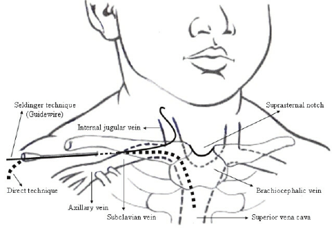
 The subclavian vein can also be cannulated using a  supraclavicular approach . This route is preferred by some authors. The essential landmark for the supraclavicular approach is the junction of the lateral border of the clavicular head of the sternocleidomastoid with the clavicle. The point of cutaneous puncture lies 1 cm superior and 1 cm lateral to this junction. The junction of the sternocleidomastoid with the clavicle defines the claviculosternomastoid angle.      The cannulating needle tip is angled posteriorly 5°–15° off a coronal plane and advanced along a line that bisects the claviculosternomastoid angle. This will lead to subclavian venipuncture between the clavicle and the anterior scalene muscle. Others suggest cutaneous puncture directly at the claviculosternomastoid angle and advancing the needle along the claviculosternomastoid angle bisector parallel and inferior to the clavicle to enter the vein at an insertion depth of 1–2 cm. ...

Internal Jugular Vein Approaches

Image
Points of possible catheterization: ANTERIOR CENTRAL POSTERIOR SUPRACLAVICULAR Traditional and safe approach is CENTRAL (pay attention to the direction of the needle): The patient is placed in the Trendelenburg position with his or her head turned to the contralateral side. The physician stands above the patient on the contralateral side of the bed, and a large skin wheal is raised with local anesthetic over the junction of the sternal and clavicular divisions of the sternocleidomastoid muscle.     While the medially located carotid artery (which courses under the sternal division) is palpated, a 11/2 inch, 22-gauge needle and syringe are used to locate the internal jugular vein which lies lateral to the carotid artery, immediately beneath the medial border of the clavicular division. The needle should enter the skin at a 30 to 45 angle directed laterally toward the midclavicle, thereby avoiding possible puncture of the c...