Posts

Showing posts matching the search for pe

Venous Thromboembolism Clinical Practice Guidelines (ASH, 2020)

Image
  Strong Recommendations For patients with PE ( pulmonary embolism ) and hemodynamic compromise, it is recommended that thrombolytic therapy followed by anticoagulation be used over anticoagulation alone. For patients with DVT ( deep vein thrombosis ) and/or PE who have completed primary treatment and will continue vitamin K antagonist (VKA) therapy as secondary prevention, it is recommended that an international normalized ratio (INR) range of 2.0 to 3.0 be used over a lower INR range . For patients with a recurrent unprovoked DVT and/or PE , indefinite antithrombotic therapy is recommended over stopping anticoagulation after completion of primary treatment.
Clinical presentation of Acute Pulmonary Embolism

Image
The clinical signs and symptoms of acute PE are non-specific. In most cases, Pulmonary Embolism  is suspected in a patient with dyspnoea, chest pain, pre-syncope or syncope, or haemoptysis. Haemodynamic instability is a rare but important form of clinical presentation, as it indicates central or extensive PE with severely reduced haemodynamic reserve. Syncope may occur, and is associated with a higher prevalence of haemodynamic instability and RV dysfunction.  Conversely, and according to the results of a recent study, acute PE may be a frequent finding in patients presenting with syncope (17%), even in the presence of an alternative explanation.

D-dimer Test. Elevated D-dimer level. D-dimer Range in Pragnancy

Image
D-dimer is a breakdown product of fibrin protein, which is present in the blood after the destruction of a blood clot. The products are formed during the lysis of a blood clot under the influence of plasmin and some fibrinolytics. In a number of pathological conditions characterized by the activation of blood coagulation, a constant process of formation of a fibrin clot occurs in the body, and then its splitting. 

Lung Ultrasound & the BLUE Protocol: The New "Visual" Stethoscope for Anesthesiologists

Image
In the high-stakes environment of the OR and the ICU, time is the ultimate variable. When a patient develops acute respiratory failure, waiting for a portable X-ray or transporting a hemodynamically unstable patient to CT can be risky, if not impossible. Enter the BLUE Protocol (Bedside Lung Ultrasound in Emergency) . Developed by Professor Daniel Lichtenstein, this diagnostic algorithm allows an anesthesiologist to identify the cause of dyspnea with over 90% accuracy in under 3 minutes. What is the BLUE Protocol? The BLUE Protocol is a standardized ultrasound algorithm designed for the rapid differential diagnosis of acute respiratory distress. By focusing on specific artifacts rather than traditional imaging, it provides immediate clinical answers. Standardized Scanning: The BLUE-Points To ensure reproducibility—much like the six leads of an ECG—the BLUE-protocol utilizes six standardized points (three on each side) that maximize lung coverage while avoiding the heart:

CARDIOGENIC SHOCK

Image
1 Give supplemental oxygen to achieve SpO2 ≥ 90%. 2 Intubate if SpO2 not maintained or altered mental state. 3 Get a 12-lead ECG and screen for STEMI equivalents using right side and posterior leads as appropriate.

Mechanism of hemodynamic instability in case of Pulmonary Embolism.

Image
    Patients with hypotension and shock without other cause are considered to have massive Pulmonary Embolism (PE) .     When obstruction occurs by embolism, the RV experiences an acute rise in pressure and volume, causing shift of the interventricular septum toward the left ventricle. In addition, as RV output  decreases,

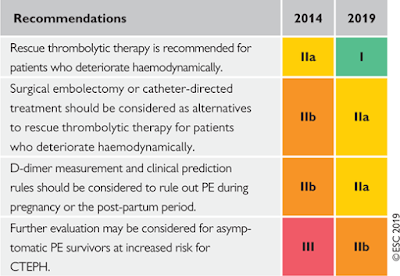
Acute Pulmonary Embolism. Changes in recommendations 2014–19

Image
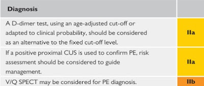
What is new in the 2019 Guidelines?

Approach to a patient with low systemic arterial blood pressure.

Image

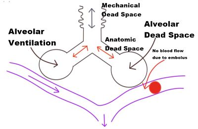
Dead space of the respiratory system

Image
  Dead space is the volume of inspired air that takes no part in gas exchange. Divided into 2 parts: ◗ anatomical dead space : mouth, nose, pharynx and large airways not lined with respiratory epithelium.  ◗ alveolar dead space :  unperfused alveoli . V entilated lung normally contributing to gas exchange, but not doing so because of impaired perfusion. Thus represents one extreme of V̇/Q̇ mismatch .

Practice Guidelines for Acute Pain Management in the Perioperative Setting: An Updated Report by the American Society of Anesthesiologists Task Force on Acute Pain Management

Image
What other guideline statements are available on this topic? These Practice Guidelines update the “Practice Guidelines for Acute Pain Management in the Perioperative Setting,” adopted by the ASA in 2003 and published in 2004.* Why was this guideline developed? In October 2010, the Committee on Standards and Practice Parameters elected to collect new evidence to determine whether recommendations in the existing Practice Guideline were supported by current evidence. How does this statement differ from existing guidelines? New evidence presented includes an updated evaluation of scientific literature and findings from surveys of experts and randomly selected ASA members. The new findings did not necessitate a change in recommendations. Why does this statement differ from existing guidelines? The ASA guidelines differ from the existing guidelines because they provide new evidence obtained from recent scientific literature as well as findings from new surveys...

Practice Guidelines for Central Venous Access: A Report by the American Society of Anesthesiologists Task Force on Central Venous Access

Image
What other guideline statements are available on this topic? Several major organizations have produced practice guidelines on central venous access  128   –   132   Why was this Guideline developed? The ASA has created this new Practice Guideline to provide updated recommendations on some issues