Posts

Showing posts from August, 2015

CABG in Patients With Acute MI

CABG in Patients With Acute MI: Recommendations CLASS I 1. Emergency CABG is recommended in patients with acute MI in whom 1) primary PCI has failed or cannot be performed, 2) coronary anatomy is suitable for CABG, and 3) persistent ischemia of a significant area of myocardium at rest and/or hemodynamic instability refractory to nonsurgical therapy is present. (Level of Evidence: B) 2. Emergency CABG is recommended in patients undergoing surgical repair of a postinfarction mechanical complication of MI, such as ventricular septal rupture, mitral valve insufficiency because of papillary muscle infarction and/or rupture, or free wall rupture. (Level of Evidence: B) 3. Emergency CABG is recommended in patients with cardiogenic shock and who are suitable for CABG irrespective of the time interval from MI to onset of shock and time from MI to CABG. (Level of Evidence: B) 4. Emergency CABG is recommended in patients with life-threatening ventricular arrhythmias (believ

Postoperative (CABG) Antiplatelet Therapy

Image
Postoperative   (CABG)  Antiplatelet Therapy: Recommendations CLASS I 1. If aspirin (100 mg to 325 mg daily) was not initiated preoperatively, it should be initiated within 6 hours postoperatively and then continued indefinitely to reduce the occurrence of SVG closure and adverse cardiovascular events. (Level of Evidence: A) CLASS IIa 1. For patients undergoing CABG, clopidogrel 75 mg daily is a reasonable alternative in patients who are intolerant of or allergic to aspirin. (Level of Evidence: C)                  AND from 2011 ACCF/AHA CABG Guideline

Bypass Graft Conduit: Recommendations

Image
Bypass Graft Conduit: Recommendations CLASS I 1. If possible, the left internal mammary artery (LIMA) should be used to bypass the left anterior descending (LAD) artery when bypass of the LAD artery is indicated. (Level of Evidence: B) CLASS IIa 1. The right internal mammary artery (IMA) is probably indicated to bypass the LAD artery when the LIMA is unavailable or unsuitable as a bypass conduit. (Level of Evidence: C) 2. When anatomically and clinically suitable, use of a second IMA to graft the left circumflex or right coronary artery (when critically stenosed and perfusing LV myocardium) is reasonable to improve the likelihood of survival and to decrease reintervention. (Level of Evidence: B) CLASS IIb 1. Complete arterial revascularization may be reasonable in patients less than or equal to 60 years of age with few or no comorbidities. (Level of Evidence: C) 2. Arterial grafting of the right coronary artery may be reasonable when a critical ( 9

Activated clotting time (ACT) vs aPTT

Activated clotting time  ( ACT ), also known as  activated coagulation time  is a test of  coagulation . [ 1] The ACT test can be used to monitor anticoagulation effects, such as high-dose  heparin  before, during, and shortly after procedures that require intense anticoagulant  administration, such as  cardiac bypass ,  cardiac angioplasty ,  thrombolysis ,  extra-corporeal membrane oxygenation  (ECMO) and continuous  dialysis . [ 1] It measures the seconds needed for whole blood to clot upon exposure to an activator of an intrinsic pathway by the addition of factor XII activators.  It is ordered in situations where the  PTT test  is not clinically useful (i.e., high-dose heparin therapy or presence of lupus anticoagulant). The ACT test is sometimes used, along with the PTT, to monitor the therapeutic effect of a direct thrombin inhibitor, such as argatroban or bivalirudin.  The ACT is measured in seconds: the longer the time to clot, the higher the degree of clotting inhibiti

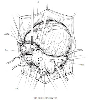
Positions of sites for cannulae and catheters.

Image
Positions of sites for cannulae and catheters Kirklin

Mitral regurgitation, perioperative hemodynamic goals

Mitral regurgitation` Heart rate/rhythm  Low heart rates are deleterious: 80-100 beats/min (sinus rhythm) ideal range  Patients often present with atrial fibrillation Preload/contractility  Maintain preload and contractility Systemic and pulmonary vascular resistance Decrease in left ventricular afterload (decrease in systemic vascular resistance) can reduce the degree of regurgitation through the valve Pulmonary hypertension may occur with chronic mitral regurgitation (often combined with mitral stenosis) and require pharmacological treatment

Mitral stenosis, perioperative hemodynamic goals

Mitral stenosis Heart rate/rhythm  Ideal heart rate 70-90 beats/min  Many patients in atrial fibrillation at time of surgery - important to control ventricular rate in this situation Preload/contractility  Maintain preload and contractility  Inotropic therapy often required following surgery (avoid tachycardia) Systemic and pulmonary vascular resistance  Long-standing mitral stenosis can lead to pulmonary hypertension  May require pulmonary vasodilator therapy postoperatively and prolonged weaning from ventilation  Inhaled nitric oxide therapy has been used to wean patients from ventilation

Preload

Image
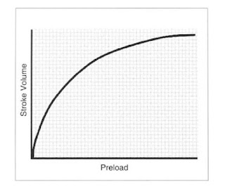
Preload represents the stretch on the muscle just prior to its contraction. Preload in the intact heart is  influenced by a number of factors that impact on ventricular filling (and hence end-diastolic stretch). These factors include:  blood volume,  venous capacitance,  ventricular compliance,  atrial function,  valvular function,    heart rate. The Frank-Starling relationship demonstrates increasing stroke volume with increasing preload. Notice that stroke volume is more sensitive to changes in preload at low values compared to high values: How do we estimate preload clinically? With the widespread use of transesophageal echocardiography (TEE), it is possible to get a reasonable estimate of chamber size and, hence, preload. In the absence of TEE, we substitute the pressure at end-diastole with the assumption that the higher the pressure, the greater the size and the greater the stretch on the contractile elements . EDP can be assessed clinically

Oxygen delivery and consumption calculations

Oxygen delivery and consumption calculations should be utilised to evaluate and optimize gas exchange: Oxygen Delivery: DO2 = 10 x CI x CaO 2 Oxygen Consumption: VO 2  = 10 x CI x (CaO2 — CvO 2 ) Where: CaO 2  (arterial oxygen content) = (Hb x 1.36 x SaO 2 ) + (0.0031 x PaO 2 ),  and  CvO 2  (mixed venous oxygen content) = (Hb x 1.36 x SvO 2 ) + (0.0031 x PvO 2 ) CI = cardiac index HB = hemoglobin SaO 2  = arterial oxygen saturation PaO 2  = partial pressure of oxygen in arterial blood SvO 2  = venous oxygen saturation PvO 2  = partial pressure of oxygen in venous blood

Cardiopulmonary Bypass Prime Solution Additives.

Image
Use of an osmotic diuretic may be advisable. Mannitol  (≈0.5 g · kg−1), a pure osmotic diuretic, can be included as part of the prime. Mannitol also has the advantage of being an effective agent against oxygen free radicals generated during CPB. Incorporating furosemide in the pump prime is practiced by many groups. It may be more advantageous to give it as a bolus in a dose of 1 to 2 mg · kg−1 at the start of rewarming, either after an interval of circulatory arrest or moderately or deeply hypothermic CPB. The short-acting adrenergic α-receptor blocking agent  phentolamine is capable of antagonizing the vasoconstriction produced by catecholamines and has been shown to produce more uniform body cooling and rewarming and improved tissue perfusion when given during CPB.B52 A bolus of 0.2 mg· kg−1 is administered just after the start of CPB and the initiation of cooling. Methylprednisolone in a single dose of 30 mg · kg−1 or  dexamethasone in a single doseof

Переливание разногрупной крови

Image
В протоколе переливания крови р. Белорусь четко указано, какую разногрупную кровь можно переливать пациентам в исключительных случаях. ( инструкция р егистрационный № 118–1103 ). Клик для увеличения:

Интерпретация результатов тестов АЧТВ и ПВ при кровоточивости у пациентов

Image

How to predict Hematocrit before starting Cardiopulmonary bypass (CPB)

You should know patient's blood volume : VpB = 1000 *f*wt where f is the proportion of body weight attributable to blood volume; f = 0.08 for infants and children up to 12 years of age, f = 0.065 for older patients. For example VpB = 1000 *0.065*80 = 5200 ml. ___________________________________________________ Than you shoud calculate the volume of patient's red cells: VpRBC = VpB * HCTp (patient's hematocrit) so : VpRBC = 5200 ml * 0.35 = 1820 ml ___________________________________________________ And finally calculation of hematocrit at the beginning of CPB : HCTpm = VpRBC / (VpB + VmB) where VmB is priming volume of CPB machine: HCT pm = 1820 / (5200 + 1300) = 0.28

Aprotinin, that is a question

" The former use of aprotinin affected the concepts of heparinization for CPB for intracardiac surgery, because this agent prolongs both clotting time and ACT, depending on the method of measurement. If aprotinin is used, an optimal recommendation is to administer the usual initial dose of heparin and add additional heparin to maintain the ACT above 700 seconds if the activating agent is Celite (diatomaceous earth). Kaolin is a more dependable activating agent, giving ACTs in the presence of aprotinin similar to those without aprotinin in vitro and during CPB. Therefore, a preferable method when aprotinin is used during CPB is to use kaolin as the activating agent and maintain the usual ACT at 480 seconds. Alternatively, the heparin concentration is measured at intervals and kept above 3 mg · kg−1. Because of results of randomized trials demonstrating that aprotinin was associated with a higher risk of death after cardiac surgical procedures than other antifibrino

Perfusion Checklist

Patient ID  ______________________ Check each item when completed, sign and date. If not applicable,  draw line through.    Bold italicized items for expedited set-up. PATIENT Patient identity confirmed Procedure confirmed Blood type, antibodies confirmed Allergies checked Blood bank number confirmed Medical record number confirmed Chart reviewed STERILITY/CLEANLINESS Components checked for package integrity/expiration Equipment clean Heat exchanger(s) leak-tested PUMP Occlusion(s) set Speed controls operational Flow meter in correct direction and calibration Flow rate indicator correct for patient and/or tubing size Rollers rotate freely Pump head rotation smooth and quiet Holders secure Servoregulated connections tested ELECTRICAL Power cord(s) connection(s) secure Servoregulation connections secure Batteries charged and operational CARDIOPLEGIA System debubbled and operational System leak-free after pressurization Solution(s) checked GAS SUPPLY Gas line(s)

Blood gases. Alfa-stat and pH-stat strategies during CPB.

Maintain arterial oxygen level not higher than 250 mmHg and less than 85 mmHg. Some clinical perfusions for cardiac surgery are performed at normothermia (≈37°C) and others at various levels of hypothermia: mild (30°C-35°C), moderate (25°C-30°C), or deep ( less than 25).Therefore, it is necessary to consider the strategy for controlling PaCO2 and, indirectly, pH.     The alpha-stat strategy is based on using the pH measured at 37°C and uncorrected for the temperature of the patient’s blood, and  maintaining this level at pH 7.4. That is, the ventilation of the oxygenator is maintained at the level appropriate for a body temperature of 37°C, no matter how low the temperature. This hyperventilation during hypothermia results in a decrease in PaCO2 and an increase in pH when the values for these are corrected for the temperature of the patient’s blood. Swan and Reeves and Rahn and colleagues have all emphasized that at low temperatures, neutrality exists

Aortic regurgitation, perioperative hemodynamic goals

Image
Important essentials Anesthesiologist should know about Aortic  regurgitation : Etiology:  Rheumatic fever Infective endocarditis Ankylosing spondylitis Takayasu arteritis Marfan’s syndrome Degenerative aortic valve disease Syphilis etc. Pathophysiology:      Aortic regurgitation produces volume overload of the left ventricle. The effective forward SV is reduced because of backward flow of blood into the left ventricle during diastole. With chronic aortic regurgitation, the left ventricle progressively dilates and undergoes eccentric hypertrophy. Eventually, as ventricular function deteriorates, the ejection fraction declines. Acute aortic regurgitation typically presents as the sudden onset of pulmonary edema and hypotension, whereas chronic regurgitation usually presents insidiously as congestive heart failure. Clinical Signs: Dyspnea on exertion, Orthopnea,  Paroxysmal nocturnal dyspnea Palpitations Angina pectoris C

Dr. John H. Gibbon

Image
John Gibbon , with his pioneering experimental work at Massachusetts General Hospital in Boston in  the late 1930s, was a major contributor to development of cardio-pulmonary bypass (CPB). One of his supporter-colleagues was a researcher at Harvard, Mary “Maly” Hopkinson. They married and continued working together, mostly at the University of Pennsylvania ’s research laboratories. By 1939, they published results of total body perfusion experiments on a number of laboratory cats that survived by employing the early apparatus invented by Gibbon.     In 1953, he performed the first successful  operation in which the patient was totally supported by CPB  when he repaired an atrial septal defect in a young woman  using a pump-oxygenator. Unfortunately, his subsequent  four patients died of a variety of problems, and he became  discouraged with the method.     The Mayo Clinic, which probably had more cardiac cases than anywhere in the U.S. , asked for pla

Monitoring for cardiac anaesthesia

Image
Essential  ECG (5-lead)  Invasive arterial pressure  Non-invasive arterial pressure  Central venous pressure  Core temperature  Urine output  Anaesthetic agent analysis  Ventilation:          - Tidal volume/respiratory rate         - Airway pressure         - End-tidal carbon dioxide   Oxygenation         -  Inspired/expired oxygen          - Pulse oximetry Optional  Pulmonary artery catheter  Transoesophageal echocardiography  Cardiac output  Oesophageal Doppler  Pulse-contour analysis  Near infrared spectroscopy  Bispectral index monitoring (BIS)

Aortic stenosis, perioperative hemodynamic goals

Image
Important essentials Anesthesiologist should know about Aortic Stenosis: N ormal aortic valve   orifice is 2.5 - 3.5 cm² Critical aortic stenosis is 0.5–0. 7 cm² A decreased valvular area causes increased pressure gradient (  mmHg ):  Mild  <  25 Moderate 25 - 40 Severe  >  40 Very severe  >  70 Triad of Symptoms:  Dyspnea on exertion Angina Orthostatic or exertional syncope Diastolic dysfunction is the result of an increase inventricular muscle mass, fibrosis, or myocardial ischemia. Loss of atrial systole can precipitate congestive heart failure or hypotension in patients with aortic stenosis. Patients may experience angina even in the absence of Coronary Artery Deasease. Once symptoms develop, most patients, without surgical treatment, will die within 2–5 years.  ANESTHETIC MANAGEMENT click to enlarge:

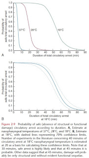
Probability of safe circulatory arrest

Image
from Kirklin