Posts

Showing posts from October, 2021

Mechanical Complications of Venous Catheterization and their Countermeasures
Image
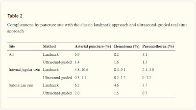
  Arterial puncture and hematoma Central venous catheterization rarely accompanies the formation of hematoma. However, with the jugular vein, and particularly when the carotid artery is mistakenly punctured, the formation of a hematoma may block the upper airway. If an artery is mistakenly punctured during SV puncture, external compression to stop the bleeding can be difficult to apply. If a catheter of 7 Fr (equivalent to a diameter of 2.3 mm) or less is inserted into an area, where compression is possible and the catheter can be withdrawn and external compression applied for 10 min, then it can be withdrawn with no problem. In other words, if an artery is mistakenly punctured by a needle thinner than 14 G (equivalent to a diameter of 2.1 mm), hemostasis may be possible via compression. However, if a catheter or dilator larger than 7 Fr is inserted into an artery or a vessel for which compression is not possible, a cardiovascular surgeon should be brought into withdraw the catheter sa

Confirmation Methods for Central Venous Catheterization

Image
  The spread of ultrasound devices has reduced the frequency of mechanical complications. However, critical situations can arise when mechanical complications occur. In a 2004 report, the central venous catheter-related mechanical complications with the highest mortality rates were pulmonary artery damage, hemothorax, cardiac tamponade , and air embolism, in that order .

Selection of Intravenous Catheters and Sites

Image
  Peripheral and Midline Catheter Recommendations 1. In adults, use an upper-extremity site for catheter insertion. Replace a catheter inserted in a lower extremity site to an upper extremity site as soon as possible. Category II  2. In pediatric patients, the upper or lower extremities or the scalp (in neonates or young infants) can be used as the catheter insertion site. Category II 

Optimal Central Venous Catheter Placement

Image
  Confirm that the catheter is placed correctly in the vein and that the tip is in the desired position using a chest radiograph. Ideally, the tip of the catheter should be roughly parallel with the wall of the superior vena cava, caudal to the inferior margin of the clavicle, between the third rib and the fourth/fifth thoracic vertebra, and cranial to the bifurcation of the trachea or right primary bronchus (see Fig  ).

Ultrasound-guided central venous catheterization. Long-axis in-plane technique.

Image
Ultrasound-guided central venous catheterization. Long-axis in-plane technique.   In this technique, the needle is advanced inside the ultrasound scan plane while observing the long axis of the vein . Unlike the short-axis out-of-plane, puncturing is truly performed in real time. Although this technique can be applied clinically with the Internal Jugular Vein if a small probe is used, it can be difficult to use regular-sized probes on the necks of small adults due to lack of space to manipulate. This technique is particularly useful with the IAV. With the long-axis in-plane technique, penetration of both the anterior and posterior walls of the vein does not occur very often, but the needle may penetrate the vein if the needle is directed from the anterior wall toward one of the lateral walls . Depicting the long axis of the center of the vein can prevent a needle directed toward a lateral wall from penetrating the vein  (Fig. 1 ). Fig. 1  Long-axis in-plane technique. To perform the p