Posts

Showing posts from November, 2016

Transcatheter aortic valve implantation (TAVI):Indications and Contraindications

Image
This new approach for the treatment of symptomatic patients with severe aortic stenosis (AS) has been shown to be feasible and safe in patients at very high or prohibitive surgical risk.  The following are indications for Aortic Valve Replacement (AVR) for severe Aortic Stenosis that apply to either surgical aortic valve replacement (SAVR) or transcatheter aortic valve implantation (TAVI):

TAVR heart procedure for inoperable aortic stenosis.

Image
The Transcatheter Aortic Valve Replacement (TAVR) procedure offers hope for heart patients with inoperable aortic stenosis (failing heart valves). TAVR valves are inserted via a catheter through the femoral artery, without requiring open heart surgery. 

Measures for reducing brain injury during cardiac surgery.

Image
Brain injury is a major source of patient morbidity after cardiac surgery and is associated with prolonged hospitalization, excessive operative mortality, high hospital costs, and altered quality of life. Main reasons: cerebral embolism by air, fat, atheromatous debris, thrombus (micro- or macroemboli) 50% of strokes; hypoperfusion inflammatory processes evoked from CPB and/or ischemia/reperfusion injury; Clinical forms of brain injury and relative frequencies: Clinical manifestation Frequency Stroke   Low risk patient ≤ 1%   High risk patient 5% to 16% Encephalopathy 8.4% to 32% Neurocognitive dysfunction   Hospital discharge 40% to 75%   One Month after surgery 12% to 30% Recommendations for measures to protect brain injury during cardiac surgery: see more:  Brain Protection in Cardiac Surgery

dir.ikernel.org

Глобальный каталог сайтов регистрирует сайты бесплатно!

Acceptable blood loss in cardiac surgery.

Image
 It's hard to make decision about reexporation after cardiac surgery. This table helps to identify acceptable blood loss.  But it's more difficult to divide "surgical" (from blood vessel) and "anesthetic" (coagulopathic) blood loss 😀

How to Recognize Type of Coronary Dominance.

Image
There are three types of coronary dominance. It depends on what coronary  artery supplies the   posterior descending artery . It can be estimated angiographically. First, h ave a look at the coronary anatomy. P osterior descending artery is written in red color. "RIGHT-DOMINANT" (70% of population): posterior descending artery is supplied by the right coronary artery ; "LEFT-DOMINANT" (10% of population):  posterior descending artery is supplied by the circumflex artery , a branch of the left artery; "CO-DOMINANT" (20% of population):   posterior descending artery is supplied by both the right coronary artery and the circumflex artery.        Dominance has important implications in  myocardial ischaemia and infarction.        The tissue separating the right and left ventricles of your heart (the interventricular septum) receives blood predominantly from the left coronary artery, but the exact breakdown of blood source is deter

Steps for successful insertion of a laryngeal mask.

Image
The laryngeal mask airway (LMA) is an acceptable alternative to mask anesthesia in the operating room. It is often used for short procedures when endotracheal intubation is not necessary.   Here some important steps for successful insertion of LMA   (click to enlarge) : Relative contraindications for the LMA: patients with pharyngeal pathology (eg, abscess) pharyngeal obstruction full stomachs (eg, pregnancy,hiatal hernia) low pulmonary compliance (eg, restrictive airways disease) requiring peak inspiratory pressures greater than 30 cm H2O.

Sevoflurane. Features, Organ Effects and Contraindications.

Image
    Sevoflurane used as an inhalational (volatile) anaesthetic for induction and maintenance of general anesthesia. Sevoflurane acts as a positive allosteric modulator of the GABAA receptor and an NMDA receptor antagonist .

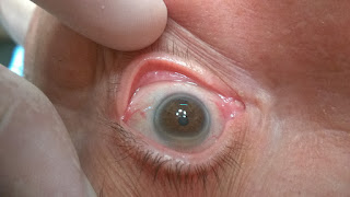
Look into the patients eyes

Image
Do not ignore physical examination of your patient. It's interesting. For example, today's case: about 70 years old male, CABG procedure. What symptom do you see? Answer is under the spoiler ) ANSWER : Arcus senilis (or arcus senilis corneae) is a white, grey, or blue opaque ring in the corneal margin (peripheral corneal opacity), or white ring in front of the periphery of the iris. It is present at birth but then fades; however, it is quite commonly present in the elderly. It can also appear earlier in life as a result of hypercholesterolemia So it is sign of diffuse atherosclerosis .

Surgeon Has Come!

Image
And good nurse will manage it easily!

Risk factors for postoperative pulmonary complications.

Image
Table of risk factors for postoperative pulmonary complications. Click to enlarge: ASA, American Society of Anesthesiologists. 1. Within each evidence category, risk factors are listed according to  strength of evidence, with the first factor listed having the strongest evidence. 2. Subsequent evidence indicates that this is a probable risk factor. Data from Smetana GW, Lawrence VA, Cornell JE, et al: Preoperative  pulmonary risk stratifi cation for noncardiothoracic surgery: systematic  review for the American College of Physicians, Ann Intern Med  2006;144(8):581-595.

Propofol Infusion Syndrome (PRIS)

Image
Propofol-related Infusion Syndrome is an acute refractory bradycardia leading to asystole and one or more of: Metabolic acidosis (base deficit > 10 mmol.l(-1)) Rhabdomyolysis Hyperlipidaemia Enlarged or fatty liver Risk Factors: Assosiated with propofol infusions at doses higher than 4 mg/kg/h  for greater than 48 h duration. Young age Low carbohydrate intake Corticosteroids intake Catecholamine infusion Pathogenesis:      Unclear, but may be associated with    impaired mitochondrial fatty acid metabolism,  mediated by propofol.   Clinical signs: Hemodynamic instability Requirement of  inotrope support Green urine ECG: right bundle branch block with convex-curved ('coved type') ST elevation in the right praecordial leads (V1 to V3). Blood gases: lactic acidosis;  Electrolytes: hyperkalaemia (in case of rhabdomyolysis, acute kidney failure) Lipaemic serum Treatment: Discontinuation of propofol infusion Haemo

Electrocardiographic Signs of Ischemia

Image
  Ischemic changes, depending on localization of Myocardium Ischemia and Infarction see below. They involve changes in T-wave morphology, including inversion, tenting, or both. More obvious ischemia may be seen in the form of progressive ST-segment depression. Down-sloping and horizontal ST depressions are of greater specificity for ischemia than is up-sloping depression.

Laryngeal Nerve Injury, Causes and Diagnostics

Image
The  laryngeal nerve  is a nerve branching down from the vagus nerve ( Cranial Nerve X) towards the larynx. It then branches into the  superior  and  recurrent laryngeal nerves. See anatomy on the picture. Main Causes of Injury: Complications of neck and chest surgery ( thyroid, lung, heart, or cervical spine surgery) A breathing tube in the airways (endotracheal tube) A viral infections that affect the nerves Tumors in the neck and upper chest, such as   thyroid   or   lung cancer Diagnostics: Do you like it?

Are you a Doctor?

Image
Are you a doctor? Every Physician sometimes hears this question in a very unpredictable circumstances... Have a good mood! Do you like it?

Hyperkalemia, Levels and ECG Changes

Image
Do you like it?

Treatment Algorithm for Duration of P2Y12 Inhibitor Therapy in Patients With Recent ACS (NSTE-ACS or STEMI)

Image
click the picture to enlarge:     Arrows at the bottom of the figure denote that the optimal duration of prolonged DAPT is not  established. Aspirin therapy is almost always continued indefinitely in patients with coronary artery disease. *High bleeding risk denotes those who have or develop a high risk of bleeding (e.g., treatment with oral anticoagulant therapy) or are at increased risk of severe bleeding complication (e.g., major intracranial surgery). ACS indicates acute coronary syndrome; BMS, bare metal stent; CABG, coronary artery bypass graft surgery; DAPT, dual antiplatelet therapy; DES, drug-eluting stent; lytic, fibrinolytic therapy; NSTE-ACS, non–ST-elevation acute coronary syndrome; PCI, percutaneous coronary intervention; and STEMI, ST-elevation myocardial infarction. Do You Like It?                               

Some Facts and Tips On Propofol

Propofol was discovered in the Biology Department at ICI Pharmaceuticals Division (now  AstraZeneca )  in the UK (1977)

Doctor's humor. Laughter is not the best medicine...

Image
Happy Friday! Time To Relax!

QUIZ 2. HEMODYNAMIC CHANGES DURING CAROTID ENDARTERECTOMY.

Image

LOCALIZATION OF CULPRIT ARTERY IN STEMI. MNEMONIC

Image

TYPES OF THE ARTERIAL WAVEFORMS

Image
Configuration of the arterial waveform at various sites in the arterial tree. (From Blitt CD, Hines RL: Monitoring in anesthesia and critical care medicine, ed 3)

ECG MNEMONIC. THE DIAGONAL RULE.

Image

Subcutaneous Engineered Stabilization Devices (ESDs)

Image

Five Key Takeaways from the New 2016 Infusion Therapy Standards of Practice

Image

True Anesthesiologist's Monitor

Image
Are you addicted to Facebook?  So, this gadget is for you!

ECG. Left Ventricular Hypertrophy.

Image
Left ventricular hypertrophy is an increase in the size of the left ventricle. When the wall of  the left ventricle increases in thickness, the heights of the QRS complexes increase. Tall voltage is usually present in Leads I , aVL , V5 and V6 . Tall voltage will be recorded in the form of tall R  waves. Likewise, when the myocardium as abnormally thickened and electrical activity takes longer to traverse throughout the whole heart, thus the duration of the QRS complex may be widened . Also, repolarization may be affected via similar mechanisms which can result in abnormal ST segments or T waves . This is referred to as "LVH with strain" or "LVH with repolarization abnormality".  The typical pattern with left ventricular hyertrophy includes deviation of the ST segment in the opposite direction of the QRS complex (discordance) and a typical T wave inversion pattern is present. ECG Criteria Sokolow-Lyon Criteria : Add the S wave in V1 plus the

Treatment Algorithm for Duration of P2Y12 Inhibitor Therapy in Patients With Stable Ischemic Heart Disease

Image
click the picture to enlarge:       Patients with a history of ACS > 1 year prior who have since remained free of recurrent ACS are considered to have transitioned to SIHD. Arrows at the bottom of the figure denote that the optimal duration of prolonged DAPT is not established. Clopidogrel is the only currently used P2Y12 inhibitor studied in patients with SIHD undergoing PCI. Aspirin therapy is almost always continued indefinitely in patients with coronary artery disease.      *High bleeding risk denotes those who have or develop a high risk of bleeding (e.g., treatment with oral anticoagulant therapy) or are at increased risk of severe bleeding complication (e.g., major intracranial surgery). ACS indicates acute coronary syndrome; BMS, bare metal stent; CABG, coronary artery bypass graft surgery; DAPT, dual antiplatelet therapy; DES, drug-eluting stent; Hx, history; MI, myocardial infarction; PCI, percutaneous coronary intervention; SIHD, stable ischemic heart disea

MNEMONIC FOR DOPAMINE DOSES

Image
B eta effects at  L ow dose A lpha effects at  H igh dose Dopaminergic effects: 0.5-2 mcg/kg/min IV  (low dose): May increase urine output and renal blood flow  Beta1 effects: 2-10 mcg/kg/min  IV (medium dose): May increase renal blood flow, cardiac output, heart rate, and cardiac contractitlity (B1-adrene) Alpha effects: >10 mcg/kg/min  IV (high dose): May increase blood pressure and stimulate vasoconstriction; may not have a beneficial effect in blood pressure; may increase risk of tachyarrhythmias

Etomidate - Heart-friendly Induction Agent with Precautions.

Image
    Etomidate is a hypnotic agent. It is a short-acting intravenous anaesthetic agent used for the induction of general anaesthesia and sedation. Of all of the available intravenous induction agents, etomidate consistently demonstrates the least direct myocardial depression in several in vitro models. Two well-designed studies using adult human atrial and ventricular tissue demonstrated no effect of etomidate on myocardial contractility in concentrations seen in clinical use.     Dosing . Initial: 0.2 to 0.6 mg/kg IV over 30-60 seconds for induction of anesthesia; maintenance: 5-20 mcg/kg/minute. Beneficial effects of etomidate are: rapid onset of action safe cardiovascular risk profile easy dosing profile slight suppression of ventilation lack of histamine liberation protection from myocardial and cerebral ischemia able to decrease intracranial pressure and maintain a normal arterial pressure in cases of traumatic brain injury  But the

EFFECTS OF MUSCLE RELAXANTS ON CARDIOVASCULAR SYSTEM

Image
click the picture to enlarge: HR        - Heart Rate SVR      - Systemic Vascular Resistanse

THORACOABDOMINAL AORTIC ANEURYSM EXTENTS.

Image
In 1986, Crawford described the first TAAA ( t horacoabdominal aortic aneurysm   ) classification scheme based on the anatomic extent of the aneurysm (click the picture to enlarge) Type I involves most of the descending thoracic aorta from the origin of the left subclavian to the suprarenal abdominal aorta. Type II is the most extensive, extending from the subclavian to the aortoiliac bifurcation. Type III involves the distal thoracic aorta to the aortoiliac bifurcation.  Type IV TAAAs are limited to the abdominal aorta below the diaphragm.  Type V , which extends from the distal thoracic aorta including the celiac and superior mesenteric origins but not the renal arteries ( Safi’s group. )

QUIZ. Allergic reaction under anaesthesia

Image
    A man is scheduled for elective noncardiac surgery. He reports collapsing due to a severe allergic reaction under anaesthesia in the past, but he is unsure which agent was the reason. There are no notes available.    Which of the following should you avoid as the most likely causative agent? (answer is at the bottom)        A. Antibiotics B. Rocuronium C. Propofol D. Ketamine E. Midazolam

CARBOXYHEMOGLOBIN. HOW TO AVOID UNDERESTIMATING COHb LEVELs

Image
    The COHb levels should also take into account the delay in sampling after removal from exposure. The half-life of COHb is 4 hours during air breathing (Douglas et al. 1912) and most patients will not have received supplemental oxygen between removal from exposure and blood sampling. A nomogram (see Figure), based on the admission COHb concentration and the time after removal from exposure, is used to obtain an approximate indication of the COHb value at exposure. By using this information, an underestimate of the level of exposure in individual patients can be avoided; and therefore where the patient has been transferred, for instance from a district general hospital to the Regional Burns Unit, with some inevitable delay in sampling, a meaningful value for COHb can be obtained. The value for COHb in the blood has also been found to be useful in identifying patients who are likely to to need intensive therapy. Thus COHb estimation may help to select high-risk patients for ele

Clinical correlation of bispectral index (BIS). Electroencephalographic (EEG) changes observed with increasing depth of anesthesia

Image
Clinical correlation of   bispectral index (BIS) (A).  Electroencephalographic (EEG) changes observed with increasing depth of anesthesia (B). (Adapted from Johansen JW, Sebel PS: Development and clinical application of electroencephalographic bispectrum monitoring. Anesthesiology 93:1336, 2000). (Redrawn From Kelley SD: Monitoring Level of Consciousness during Anesthesia and Sedation. Natick, MA, Aspect Medical Systems, 2003).

ANESTHESIOLOGIST's ABCD

Image
click to enlarge Anesthesiologist's ABCD: A-airways B - Breathing C - Chair and Coffee D - Doubt in deal with surgeons